Radiology/Medical Imaging

Home » Services » Radiology/Medical Imaging

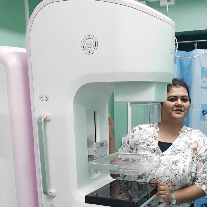
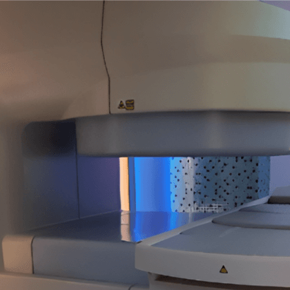
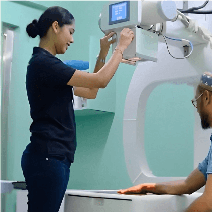
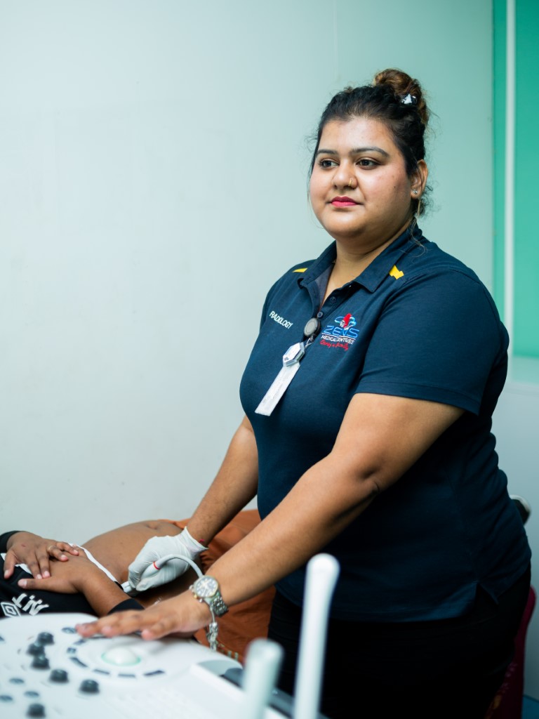
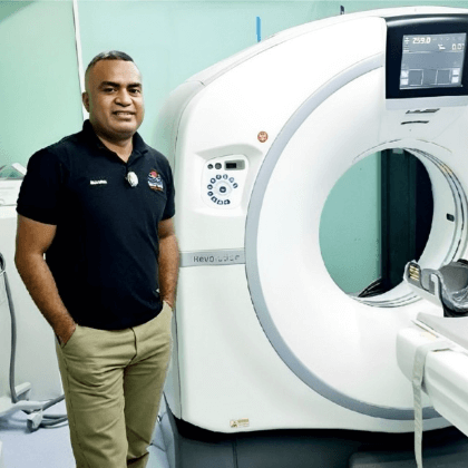
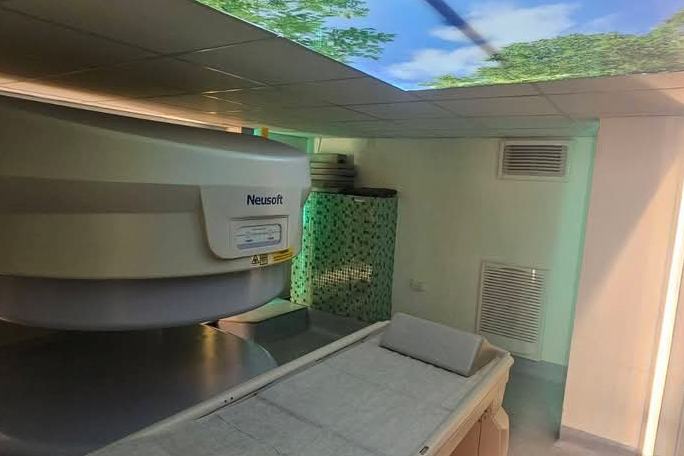
At Zens Medical Radiology, we are committed to delivering precision-driven, compassionate diagnostic imaging services that empower physicians and patients to make informed healthcare decisions. Our state-of-the-art facility combines advanced technology with a team of highly skilled radiologists and technicians, ensuring accurate and timely imaging solutions.


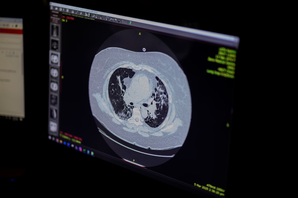
We take pride in offering a comprehensive range of services, including X-rays, CT scans, MRI, ultrasound, and specialized imaging procedures, all designed to support your healthcare journey. With a focus on patient comfort and diagnostic excellence, Zens Medical Radiology stands at the forefront of medical imaging care.

Meet Our Radiology Experts

At Zens Medical Radiology, we are led by the expertise and visionary leadership of Dr. Laitia Lolokabaira, a seasoned Radiologist with 19 years of medical experience, including 15 years dedicated to radiology with the aid of the MaxHealth Radiologists India Team.

His extensive background spans roles as Head of Radiology at Labasa Hospital and key positions in clinical advisory and quality improvement committees across major healthcare institutions in Fiji.

His unwavering commitment to excellence and patient-centered care ensures that Zens Medical Radiology provides the highest standards of diagnostic precision and healthcare solutions for our patients.

Dr. Laitia Lolokabaira

Director of Radiology Services

Imaging Department

What to expect for Ultrasound Scan

Before

You will need a referral/ request form from your doctor or physiotherapist to be able to conduct a scan.
Some ultrasound examinations need no preparation. Preparation is required for certain scans.
For early pregnancy, pelvic/ gynecological, kidney and prostate scans, your bladder needs to be full in order to optimally view your pelvic organs. The best way to achieve a full bladder is to first empty your bladder one and a half hours prior to your appointment. Then drink one liter of water (4–5 glasses) over the next half hour. Ingestion of water should be completed one hour prior to the examination. Please do not go to the toilet.
For scans of your abdomen, such as the gallbladder, it is important you do not eat or drink anything for six to eight hours beforehand.
For vascular ultrasounds, prior booking is required.
For a musculoskeletal ultrasound, generally no preparation is required.

During

On arrival, present to reception with your referral form if you or your referrer have not already sent it through. You may be given a consent form to complete. The examination usually takes 30-60 minutes depending on the area to be scanned. You may be asked to change into a gown.
Water-based gel is applied on your skin over the area to be examined. The Medical Imaging Technologist uses a transducer over the gel to scan and acquire images as required.
For pregnancy scans, as this is an important medical examination, it is best to give your Medical Imaging Technologist some time to focus on the examination before asking questions. You are not able to record your scan but ultrasound images can be copied and given to you after the scan.

After

You will be able to resume your normal activities after the ultrasound scan. Results of your scan will be sent to your doctor as soon as possible. You can also request a copy of your results once they are available.

Benefits of Ultrasound

Non-invasive: No incisions or needles are required. 
Safe: No ionizing radiation is used. 
Portable: Ultrasound equipment can be moved easily, making it suitable for a variety of settings. 
Real-time Imaging: Provides a dynamic view of internal structures and movement. 
 

Limitations of Ultrasound

Not Ideal for Bone Imaging:
Ultrasound waves don’t penetrate bone very well, so it’s not the best for imaging bone structures.
Air Interference:
Ultrasound waves can be significantly affected by air, making it difficult to image the lungs

what is (CT) Computed Tomography

CT (Computed Tomography) scans use X-rays and computer technology to create detailed, cross-sectional images of the body, revealing internal structures like bones, organs, soft tissues, and blood vessels. Unlike standard X-rays, which produce two-dimensional images, CT scans generate a series of “slices” that can be combined to create three-dimensional views. These images are valuable for diagnosing a wide range of conditions, including tumors, fractures, and internal bleeding.

What to expect for CT Scan

Before

You need a referral/ request form from your doctor to be able to book an appointment for this scan.
When you make an appointment for your scan you will be advised as to whether you require any specific preparation and we will go through the process with you then.
As a general guide you will be advised to not eat anything six to eight hours prior to your appointment if you are required to have Contrast. Contrast is a colourless fluid that is injected at the time of your scan to improve the definition of the images.
For abdominal examinations you may also be required to drink water before your scan.
All patients will be asked to present 10 minutes prior to their appointment time as you may need to complete a set of questionnaire.

During

Depending on the type of scan you are having, your appointment may take up to 60 minutes to complete.
Your Medical Imaging Technologist (MIT) will guide you through the whole process and gain consent from you. For some examinations you may be asked to change into a gown and to remove metallic objects such as jewelry and hearing aids.
The CT MIT, who is trained in the use of X-ray equipment and CT scanning, will position you on the bed and take care of you throughout the scan. The MIT will need to leave the scan room briefly whilst it is in progress but will remain in contact with you via an intercom and a viewing window.
It is important that you keep still while the scan takes place. If you have difficulty lying flat, have poorly controlled pain or suffer from claustrophobia, please discuss this with us prior to your scan.
If you need to have Contrast for your scan this is given through a small needle that is placed in your vein. Injection of Contrast may give you a brief sensation of warmth throughout the body and maybe a metallic taste in your mouth. These sensations are expected and normal and are of no cause for concern.

After

After the examination, you are able to resume your normal activities.
Results of your scan will be sent to your doctor after 3 working days – depending on the Radiologist workload. You can also request a copy of your results.

Benefits


Detailed Images: CT scans provide more detailed images than standard X-rays, allowing for better visualization of internal structures and abnormalities. 
Cross-sectional Views: CT scans produce cross-sectional images, or “slices,” of the body, allowing for a more complete assessment of internal structures. 
3D Reconstruction: CT scan data can be used to create three-dimensional images, which can be helpful for surgical planning and diagnosis. 
Speed and Efficiency: CT scans are relatively fast and can be performed quickly, making them useful in emergency situations and for certain types of cancer screening. 
 

Risk

Higher dose of radiation when compared to plain xrays

what are X-rays

X-rays are a type of electromagnetic radiation used in medical imaging to create pictures of the inside of the body. They are commonly used to diagnose conditions like fractures, tumors, and infections. X-rays work by passing radiation through the body, with different tissues absorbing varying amounts, which creates an image.

A person looking at a machineAI-generated content may be incorrect.

What to expect for X-Ray

Before

You need a referral/ request form from your doctor to be able to book an appointment for this scan.
When you make an appointment for your scan you will be advised as to whether you require any specific preparation and we will go through the process with you then.
As a general guide you will be advised to not eat anything six to eight hours prior to your appointment if you are required to have Contrast. Contrast is a colourless fluid that is injected at the time of your scan to improve the definition of the images.
For abdominal examinations you may also be required to drink water before your scan.
All patients will be asked to present 10 minutes prior to their appointment time as you may need to complete a set of questionnaire.

During

In general, you undress whatever part of your body needs examination. You may be asked to wear a gown during the exam, depending on which area is being X-rayed. You may also be asked to remove jewelry, eyeglasses, and any metal objects that can show up on the X-ray.
Once you are fully prepared, your Medical Imaging Technologist will tell you how to position your body to get clear images. The machine sends a beam of radiation through your body.
It is important to stay still while the images are being taken. This will provide the clearest images possible.

After

After the examination, you are able to resume your normal activities.
Results of your scan will be sent to your doctor as soon as possible. You can request a copy of your results as well.

What is (MRI) Magnetic Resonance Imaging?

MRI (Magnetic Resonance Imaging) is a powerful medical imaging technique that uses strong magnetic fields and radio waves to create detailed, cross-sectional images of the body’s internal structures. These images help doctors diagnose a wide range of conditions, including tumors, injuries, and diseases.

What to expect for X-Ray

Before

During

After

Benefits of MRI

Non-invasive: MRI does not require surgery or the introduction of any foreign objects into the body. 
No radiation: MRI does not use ionizing radiation, unlike X-rays or CT scans. 
Detailed images: MRI provides very detailed images of the body’s internal structures.

Risk

Dynamic Content
Claustrophobia: The confined space of the MRI scanner can trigger anxiety or claustrophobia in some individuals. 
Noise: MRI scanners can be loud, potentially causing hearing loss if not addressed.

What is Mammogram

Mammography is a crucial medical imaging technique used to detect breast cancer and other breast diseases using X-rays. It involves taking X-ray images of the breast to identify abnormalities like dense areas, unusual configurations, or microcalcifications.

A person standing next to a machineAI-generated content may be incorrect.

What to expect for Mammogram

Before

You will need a referral/ request form from your doctor or physiotherapist to be able to perform Mammogram.
If this is a routine check-up, try to book your appointment the week after your period when your breasts are less sensitive. If you have new symptoms such as a palpable lump or nipple discharge, let the booking staff know when you book your mammogram.
On the day of your examination do not wear roll-on deodorant, talcum powder or lotion on your breast as these may show up on your mammogram. It’s best to wear a two-piece outfit as you will need to undress from the waist up and you will be given a gown to wear.

During

On the day of your appointment, you will be asked a few questions that will help us in assessing your risk and interpreting your mammogram. The whole procedure takes about 20-30 minutes.
Mammography involves compressing your breast tissue for a short time to x-ray the breast. The pressure can be uncomfortable, but most women cope very well. If you find the pressure causes extreme discomfort, please tell the Medical Imaging Technologist immediately.
You may be required to have additional views or ultrasound for further assessment upon request from the Radiologist.

After

After your mammogram, you may experience some redness of the skin and in rare cases, you may experience some bruising.
Results of your Mammogram will be sent to your referring doctor as soon as possible. You can request a copy of your results as well.

Benefits

Early Detection: Mammograms can detect breast cancer at an earlier stage than physical exams, often finding lumps two to three years before they can be felt

Detection of Benign Conditions

Mammography can also help detect benign conditions like cysts and fibroids, allowing for timely management and preventing unnecessary worry.

Limitations of Mammography

False Positives: Mammograms can sometimes produce false positives, leading to unnecessary worry and additional testing.
Not All Cancers Detected: Some cancers may not be visible on mammograms due to their location within the breast or the density of breast tissue.